Direct observation of large-area strain propagation on free-standing nanomembranes
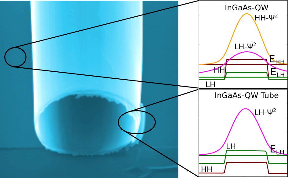
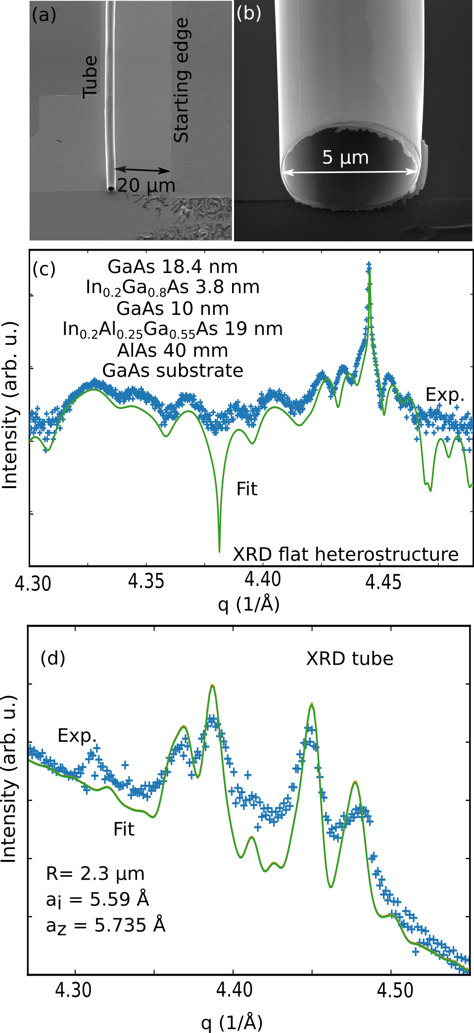
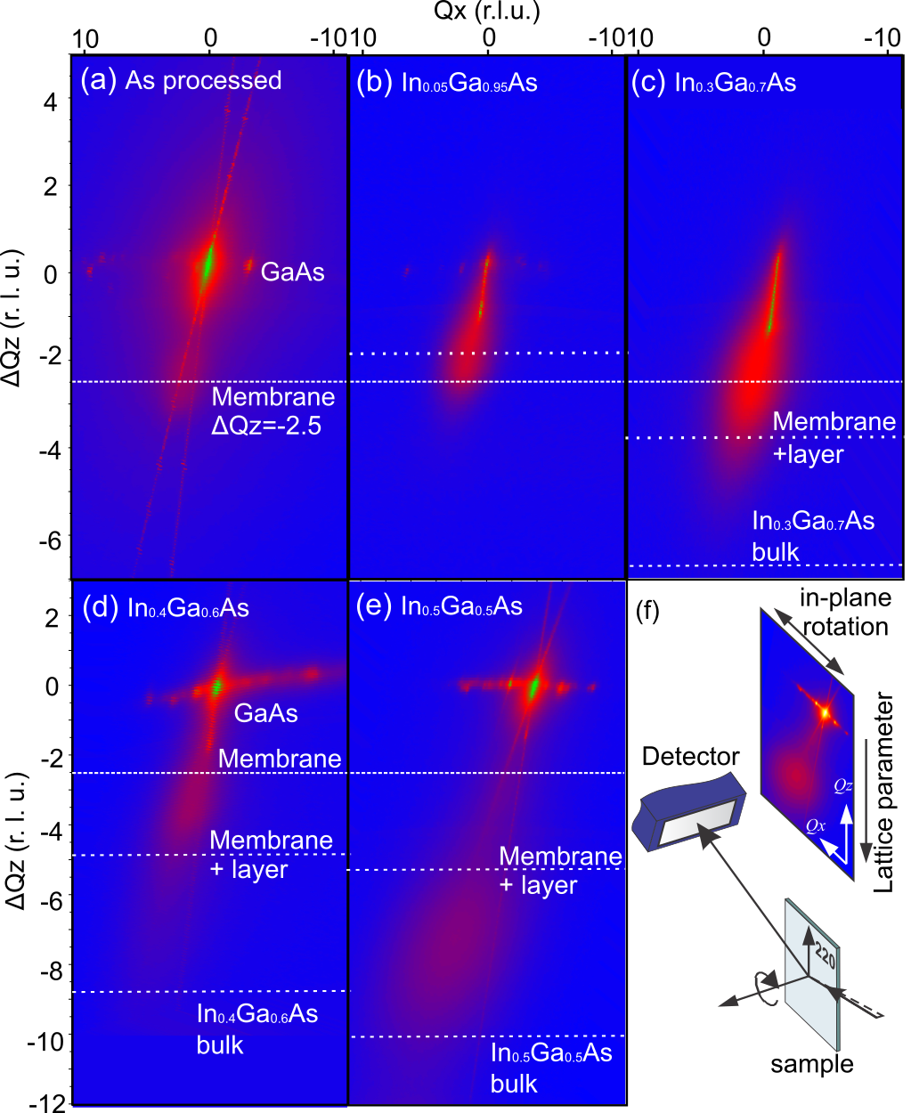
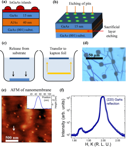
Investigations on epitaxial nanostructures with size of tens of nanometers have been a challenging issue for techniques that present high strain sensitivity but restricted spatial resolution. This is the case of recently developed x-ray nanoprobe techniques. Despite its inherent nondestructive character, submicron x-ray spots have only been successfully applied to the study of individual nanostructures which are either strain free or present extremely mild spatial lattice parameter gradients. Such limitation, with an uttermost barrier given by the diffraction limit, leads to voxel or pixel sizes between 5 and 10 nm obtained in coherent diffraction imaging or ptychographic reconstructions of real-space objects. Whenever the strain field of a nanostructure is successfully reconstructed from reciprocal space measurements, it cannot vary considerably in short distances since this would induce diffraction peak broadening and cause abrupt phase variations, leading to convergence issues on reconstruction algorithms. Here we show how epitaxial systems with large lattice mismatch and appreciable interfacial strain can be identified and directly analyzed throughout their strain field propagation in nanometer-thin crystalline membrane platforms, using the InGaAs/GaAs Stranski-Krastanov system as a model. The strain-induced footprint becomes observable along a few microns if the membrane thickness is comparable to the nanostructure size. It is possible to retrieve both interfacial strain and nanostructure size by probing individual objects.
Yuri Bernardes, Lucas A. B. Marçal, Barbara L. T. Rosa, Ailton Garcia, Jr., Christoph Deneke, Tobias U. Schülli, Marie-Ingrid Richard, and Angelo Malachias
Phys. Rev. Materials 7, 026002